
Stratasys Launches First 3D Printing Material Designed to Look Like Human Tissue on Scans

Medical professionals and device manufacturers can now create patient-specific anatomically realistic models that behave like real tissue under CT or X-ray.
Stratasys has announced the full U.S. release of RadioMatrix, a proprietary 3D printing material used with the company’s PolyJet material jetting printers, including the J5 and J5 Digital Anatomy systems. Already tested in selected pilot programs, the material is now available to American hospitals, and stands out as the first broadly available commercial 3D printing material that offers controlled, tunable radiopacity — the measure of how visible different tissues appear on X-ray and CT scan — for anatomically realistic CT-compatible models for surgical practice, pre-operative planning, and education.
3D printing is increasingly replacing cadavers and synthetic models in medical training and testing, but RadioMatrix extends that shift even further. First it moves more economical 3D printed models into the domain of imaging, offering radiologists and researchers a consistent alternative for developing and fine-tuning CT protocols without relying on human remains or standardized models.
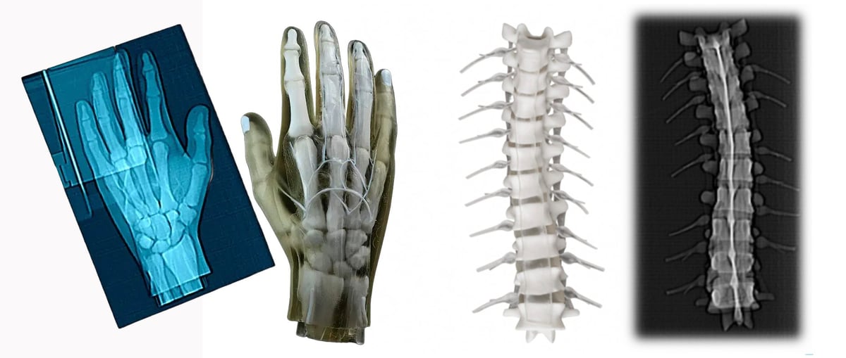
More importantly, this new materials enables highly realistic, patient-specific anatomical models that show up on scans like specific patients real tissue. Unlike traditional CT “phantoms,” which are often made from standardized materials or sourced from human cadavers, RadioMatrix enables models that can be customized, reproduced, and tuned for specific imaging needs.
“Providing full availability of RadioMatrix in the U.S. is a major step in providing cutting-edge imaging education and training,” says Erez Ben Zvi, Vice President of Healthcare at Stratasys. “By giving radiologists and device manufacturers the ability to print ultra-realistic, customized radiographically accurate models, we’re helping replace traditional phantom solutions and reliance on cadavers with customizable, repeatable, and scalable alternatives.”
Early research from the Stratasys–Siemens Healthineers collaboration shows RadioMatrix-printed phantoms can closely match real tissue on CT, with accuracy down to single Hounsfield units (HU) in areas like grey matter and veins. That level of fidelity is critical for validating new imaging techniques, optimizing scanner settings, and refining diagnostic algorithms—all while avoiding the variability and ethical concerns associated with cadaver use.
In the U.K., partners such as CPI and Beaumont Hospital are already demonstrating how radiopaque 3D-printed models can improve medical training. Their radio-realistic cerebral angiography phantoms allow clinicians to practice imaging procedures in a controlled, repeatable environment that traditional cadaver-based training cannot offer.
RadioMatrix is designed to work in combination with Stratasys’ Digital Anatomy technology, which replicates the feel and biomechanical behavior of living tissues. Together, the systems produce highly detailed, patient-specific models that preserve fine structures and pathological variations—while being entirely 3D printable and scalable for large training programs.
You May Also Like:
Listen to All3DP’s News Podcast:
License: The text of "Stratasys Launches First 3D Printing Material Designed to Look Like Human Tissue on Scans" by All3DP Pro is licensed under a Creative Commons Attribution 4.0 International License.